Radiation Therapy
LUCY
®
3-D Plus
Precision 3-D QA Phantom
Model 76-311
•
Multi-tasking radiographic and
dosimetric QA phantom
specifically developed for radiation
therapy, radiology, and stereotactic
radiosurgery
•
Verify imaging errors, compare
localization measurements of
different imaging modalities,
including CT, angiography, portal
imaging, and MRI
•
Test image distortion and slice
thickness
•
Verify target volume calculations
and CT / MRI fusion
•
Verify accuracy of treatment
planning software algorithms
•
For hardware verification, linear
accelerator gantry isocentricity,
table positioning, laser alignment,
and collimators
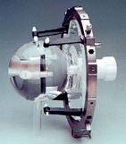
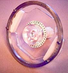
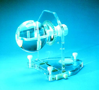
LUCY 3-D Plus sphere shown on standard alignment base
Introduction
LUCY 3-D Plus is a spherical, acrylic phantom precision-machined to simulate a patient’s
head. It contains known markers and spatial relationships which are detectable by CT,
angiography, and MRI imaging modalities, as well as various therapeutic equipment.
The LUCY 3-D Plus Phantom was developed to simulate clinical situations, verify
hardware and software components, and detect system errors.
Applications
LUCY supports all 3-D stereotactic radiotherapy and small field IMRT Systems, for
localization and treatment verification using major commercially available treatment
planning and delivery systems.
Use of the LUCY Phantom permits quantitative assessment and calibration of parameters
described in established quality assurance protocols and the commissioning of new
equipment and procedures, such as fusion and IMRT.
•
Verify planned vs. delivered dose
distribution using radiochromic film,
ion chambers, MOSFETs, TLDS,
and gel
•
Adapts to any stereotactic frame to
verify material or geometry effects
•
A commissioning tool for hardware
and software
•
A useful teaching aid
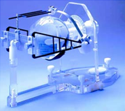
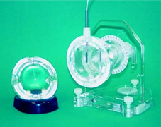
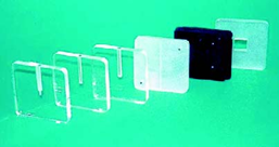
Detector in open LUCY sphere
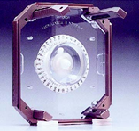
Film cassette
Multiple detector cassettes
Specifications
The LUCY phantom sphere is constructed of acrylic, a standard radiation
calibration material recommended by the AAPM Task Group 21
All screws and plugs are made of white plastic material
A 360° rotation scale is located on the vertical support, around the
sphere axis for sagital or coronal mounting
The LUCY sphere is 140 mm in diameter
Upper Hemisphere Cavity: 81 x 81 x 35 mm, for insertion of an MRI
signal generator or other 3-D volumes, e.g. stacked film or gel
container
Lower Hemisphere: has four cylindrical chambers 10 mm Ø x 25 mm
deep, for placement of radiological markers
A second cavity of 85 x 85 x 10 mm is located at the center of the
sphere, for placement of accessories and cassettes. An accessory port
of 8 mm in diameter allows for insertion of dosimetry sensors to the
center of the sphere, in the transverse mount
Weight: 11.8 lb (5.3 kg)
Available model(s)
Model
Description
76-311
LUCY 3-D Plus Precision 3-D QA Phantom,
includes one phantom sphere and one tool kit set
Alignment bases, including MRI and stereotactic frame
interfaces
Model
Description
76-311-4000
Standard Base and Clamp
76-311-4002
Stereoadapter
®
Frame Supports
76-311-4003
Load-Bearing Stereoadapter Base
76-311-4004
Brainlab Frame Interface
76-311-4005
Optional Brainlab Mask System Adapters
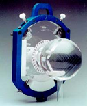
LUCY interfaced with stereotactic systems
76-311-4006
Leksell/Gamma Knife Interface
76-311-4007
Leibinger Frame Interface
76-311-4008
Radionics BRW Frame Interface
76-311-4009
Optional Radionics GTC Frame Adapters
76-311-4010
Optional Radionics MRI Frame Adapters
All OEM company trademarks are implied.
Radiological accessories
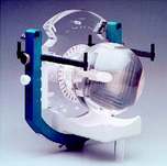
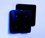
LUCY/Brainlab Interface
LUCY/Brainlab Interface Mask
System
Model Description
76-311-3006 CT Marker Cylinders, set of four
76-311-3007
Angiography Marker Cylinders, set of four
76-311-3008
MRI Marker Cylinders, set of four
76-311-3009
CT/Angio Grid Plate
76-311-3011
CT 3-Volume Chamber
76-311-3012 MRI 3-Volume Chamber
76-311-3013 Target/Treatment Verification Chamber
76-311-3021 3-D Volume Chamber with lid (supplied empty)
76-311-3022 MRI Signal Generator
Radiotherapy / dosimetry accessories
LUCY/Leksell Interface
LUCY/Leibinger Interface
Model Description
76-311-3013 Target/Treatment Verification Chamber
76-311-3014 Radiation/Optics Alignment Pointer
76-311-3030
PTW Cassette for Pin Point 0.015cm
3
(Model TN31006)
76-311-3031 IBA-Scanditronix Cassette for Chamber (Model DEB050)
76-311-3032 Exradin Cassette for chamber (Model IC-A1SL-1)
76-311-3033
IBA-Wellhofer Cassette for chamber (Model CC01) (10 mm)
76-311-3040
Radiochromic Stacked Film Assembly (85x85x10 mm)
4x2.25 mm Dividers suitable for 3x3 inch film
76-311-3041
Radiochromic Stacked Film Assembly (85x85x10 mm)
4x2.25 mm Dividers suitable for 2.5x2.5 inch film
76-311-3042
Radiochromic Stacked Film Assembly (81x81x35 mm)
14x2.25 mm Dividers suitable for 2.5x2.5 inch film
76-311-3045 MOSFET Array Cassette suitable for TLD’s
76-311-3051 B-4 Cassette for Polymer Gel Container (81x81x35 mm)
76-311-3052 B-4 Polymer Gel Container - disposable
LUCY/Radionics BRW Interface LUCY/Sandstrom Interface
References
1. R. Ramani, M.G.Ketko, P.F. O’Brien, M.L. Schwartz, “A QA Phantom for Dynamic
Stereotactic Radiosurgery: Quantitative Measurements,”
Medical Physics
, 22 (8),
(1995) 1343-1346.
2. R. Ramani, A.W. Lightstone, D.L.D. Mason, P.F. O’Brien, “The Use of Radiochronmic
Film in Treatment Verification of Dynamic Stereotactic Radiosurgery,”
Medical Physics
,
21 (3), (1994) 389-392.
76-311-4001
GE MRI Head Coil Base